An Introduction to Veterinary Radiography
Dental radiographs are essential for the proper diagnosis of oral pathology in our dog and cat patients.
By Denise Rollings, AAS, CVT, VTS (Dentistry), Key Account Manager, iM3 USA | 2025
Many oral and dental diseases cause inflammation and discomfort for our patients, even if they do show outward signs. Most of the pathology is below the gingival margin, making it difficult or impossible to see on an oral examination alone.
Types of Intraoral Radiography Equipment
Many oral and dental diseases cause inflammation and discomfort for our patients, even if they do show outward signs. Most of the pathology is below the gingival margin, making it difficult or impossible to see on an oral examination alone.
The X-Ray Generator Tube Head & Technical Adjustments
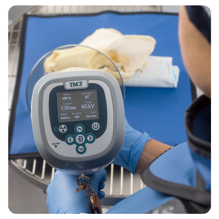
The tube head is the position-indicating device (PID) and may also be called the “cone.” The PID is what is aimed at the area of the mouth to be imaged. The exposure time, voltage (kVp), and milliamperage (mA) may be adjustable depending on the generator using the control panel.
The control panel has preset settings based on the size of the patient and the area of the mouth being imaged to help produce the best exposure for that image.
Different Types of Generators
Generators may be mounted on stands (trolley), walls, or be handheld. A wall or ceiling-mounted system requires that the station where the dental radiographs will be obtained is within reach. The trolley-mounted system may be moved to different stations. The use of a trolley system may be prohibited by the floor space of a clinic. A handheld generator needs the least amount of space and may be moved to different stations for use. State laws vary regarding generator use and must be followed.
Dental X-ray Films
Dental X-ray films are much less common today but are still (rarely) in use. In this method, the film is placed into a protective cover and, after exposure, is developed in a small chairside developer using chemicals. This process can take several minutes, and the resulting image is evaluated using a light source behind the film. While familiar to some practitioners, the system is slow and requires chemical maintenance.
Direct Digital Imaging (DR)
Direct digital imaging (DR) uses a hard sensor connected to a laptop or generator via a cord. The sensors typically come in sizes 0, 1 and 2, with size 2 being the most common. A few companies, such as iM3, offer a size 4, and a size 6 digital sensor. Due to the high cost of sensors, most facilities only purchase one size. Once exposed, the image is transferred to the computer and viewable within seconds. DR systems are the fastest and require the least radiation, but they may be limited to a size 2 sensor, meaning larger patients require multiple images for a single tooth or larger area of the mouth unless a size 4 or 6 digital sensor is used.
Indirect or computed digital imaging (CR)
Indirect or computed digital imaging (CR) uses reusable phosphor plates, which are available in sizes 0 through 6, depending on the manufacturer. The plate is placed in a protective sheath before positioning in the patient’s mouth. After exposure, the sheath is removed, and the plate is inserted into a scanner. The scanner converts the data into a digital image, erases the plate, and allows it to be reused.
This system can develop an image in as fast as 6 to 8 seconds depending on the manufacturers and settings. A CR system requires a supply of protective sheaths. CR plates are thin, flexible and available in larger sizes, making them comfortable for patients and offers the ability to obtain full mouth radiographs on a large-breed dog patient in as few as six images.

The iM3 CR 7 2.0 (left) processes one phosphor plate at a time, while the iM3 CR 8 (right) processes two simultaneously to help save time.
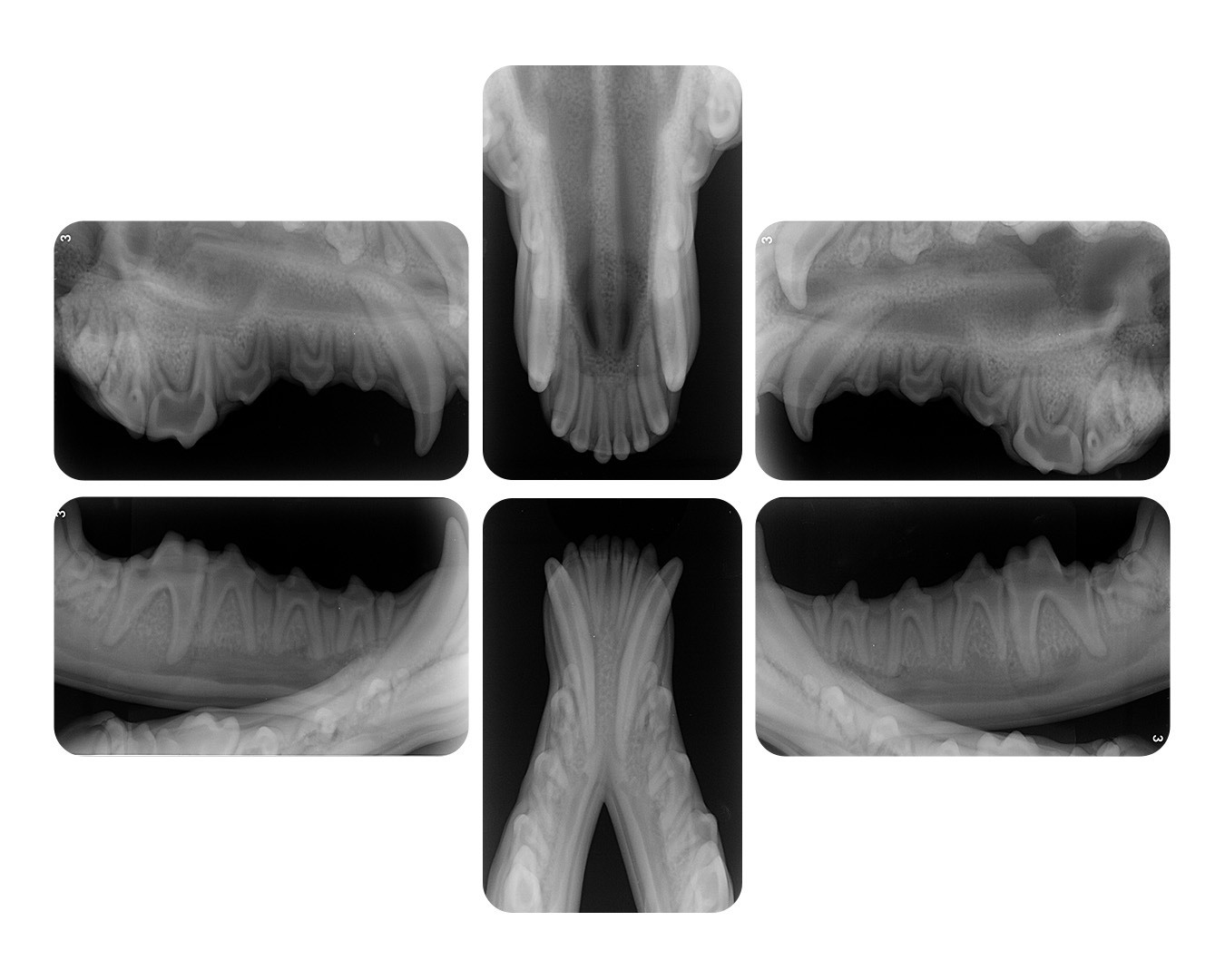
full-mouth dental X-Ray images taken on a large dog using a size 5 CR plate

Dental Imaging Software
Both DR and CR systems work with dental imaging software that allows the user to adjust brightness, contrast, rotation, inversion and magnification. Areas of the radiograph may also be circled, and pathology may be pointed out to the patient’s owner.
Most dental radiograph software systems allow the user to use full-mouth templates. These templates are preset with the order in which the user would like to obtain their dental radiographs and reduce the amount of computer time needed. These systems vary in cost, upkeep and features. It is important to research each system’s cost, maintenance needs, technical support, warranties and training options before a purchase is made.